Figure 9.

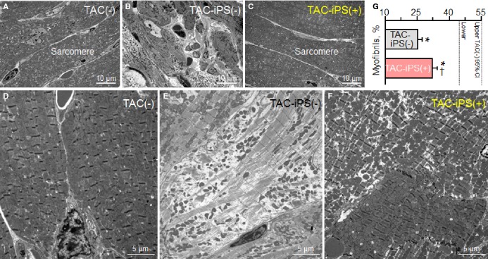
Protection of myofibrillar ultrastructure. Sarcomere ultrastructure at low- (A through C) and high-magnification (D through F) electron microscopy. Kir6.2-deficient left ventricles after 3 months of TAC demonstrated severe fibrosis [TAC-iPS(−); B] and lacked myofibrils (E). Cell-treated Kir6.2-deficient ventricles [TAC-iPS(+)] exhibited normal sarcomeres (C) with significant increase in myofibrils (F and G). *P<0.05 vs. TAC(−); †P<0.05 vs. TAC-iPS(−). CI indicates confidence interval; iPS, induced pluripotent stem; TAC, transverse aortic constriction.
