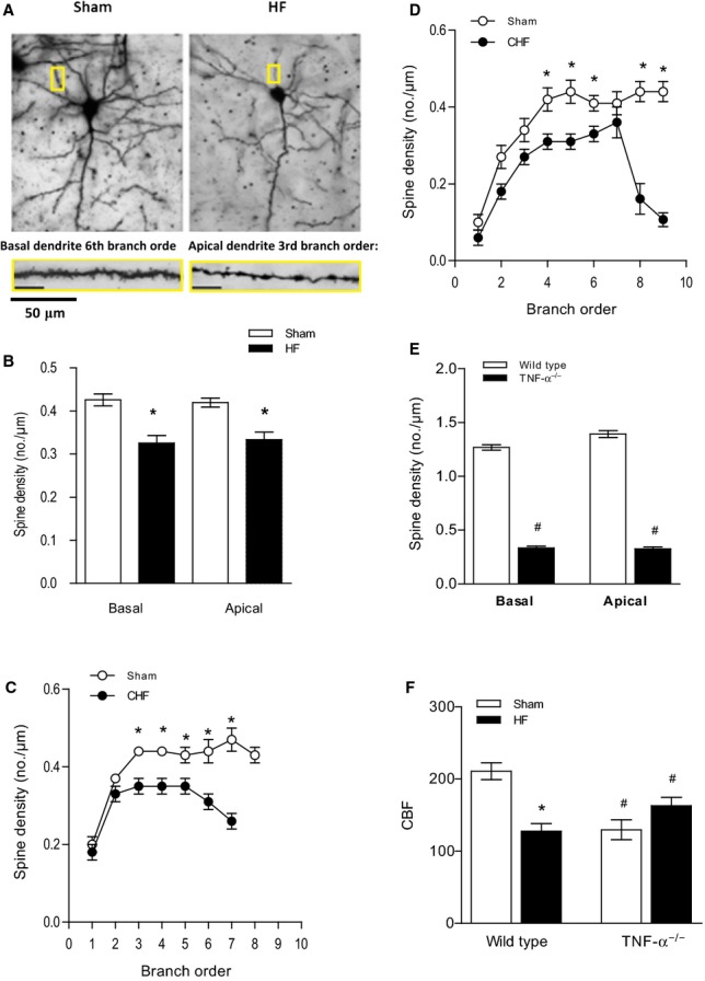
Figure 5.

HF-induced effects in the brain are ameliorated in TNF-α−/− mice. A, Representative images of dendritic spines of pyramidal cells from TNF-α−/− mice. The magnification shows details of both basal and apical dendrite morphology (5 to 7 cells per animal). B, Total spine density of basal and apical dendrites was significantly reduced in HF mice (4 to 6 cells per animal). Breakdown analysis of spine density found that significant reductions occurred across several branch orders of (C) basal dendrites and (D) apical dendrites between sham and HF mice. D, The average spine density of TNF-α−/− sham mice was significantly lower than that of normal wild-type animals in both basal and apical dendrites. For all tasks: sham n=24, HF n=24. *P<0.05, #P<0.001. E, HF had no effect on cerebral blood flow in TNF-α−/− mice (n=5, P=0.9069) compared with TNF-α−/− sham-operated mice. F, CBF, shown as mL/(100 g×minute), was significantly reduced compared with wild-type control animals (n=5, P<0.001). *P<0.05 within a group, #P<0.05 between wild-type and TNF-α−/− mice. CBF indicates cerebral blood flow; CHF, congestive heart failure; HF, heart failure; TNF-α, tumor necrosis factor-α.
