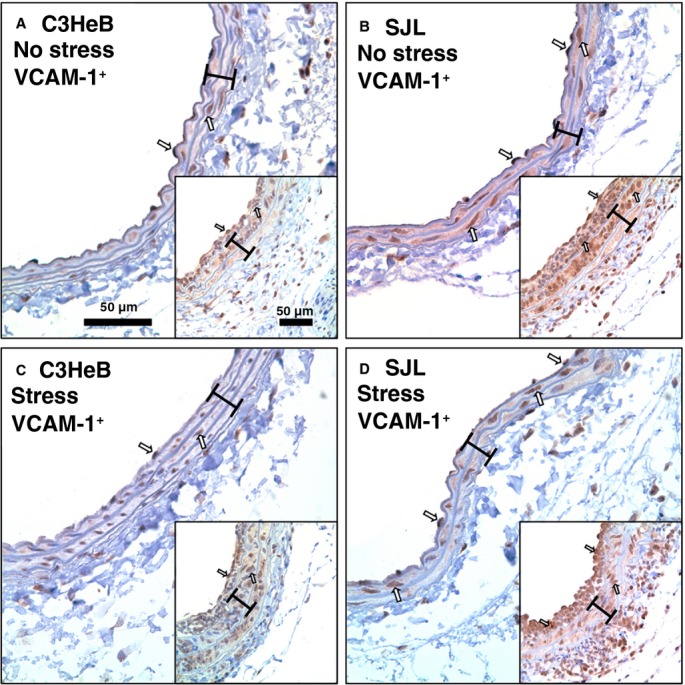
Figure 3.

Carotid inflammation in mouse strains with or without stress. Representative vascular cell adhesion molecule 1 (VCAM-1)+ staining of noninjured carotids: A, C3HeB mice, no stress. B, SJL mice, no stress. C, C3HeB mice, with stress. D, SJL mice, with stress. Insets show VCAM-1 immunoreactivity in carotid arteries after the injury across respective groups (A through D). VCAM-1+ cells are stained dark brown (open arrows). Brackets show area between internal and external elastic lamina. Magnification bar is 50 μm.
