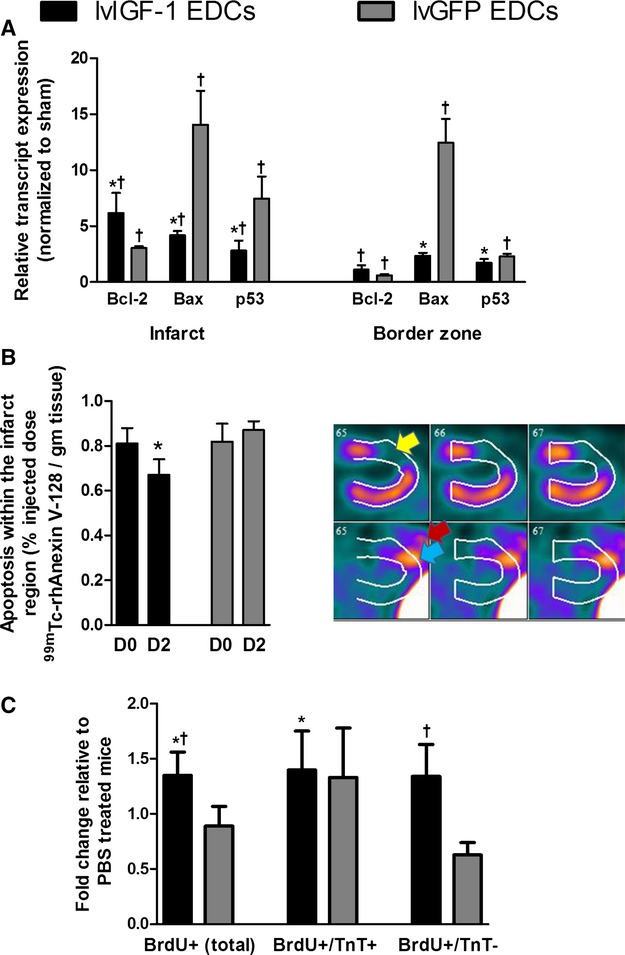
Figure 7.

Transplantation of human EDCs genetically engineered to overexpress IGF-1 reduces apoptosis and promotes endogenous regeneration. A, Quantitative polymerase chain reaction of microdissected infarct and peri-infarct areas from mice treated with lvIGF-1– or lvGFP-transduced EDCs demonstrating reduced expression of apoptotic markers 7 days after EDC injection. *P≤0.05 vs lvGFP-transduced EDCs, †P≤0.05 vs sham-treated animals; n=4 mice with 3 technical repeats. B, Single-photon emission computed tomography quantification of apoptosis within the 201Tl-defined infarct region before (D0) and 2 days after injection of lvIGF-1– or lvGFP-transduced EDCs (D2). Also shown are representative horizontal long-axis images from a mouse 2 days after injection of lvIGF-1 demonstrating 201Tl perfusion images (upper panel) with myocardial infarction (yellow arrow) and 99mTc-rhAnnexin V-128 images (lower panel) with uptake in the area of infarction (blue arrow) and surgical site (red arrow). *P≤0.05 D2 vs D0; n=3 mice. C, NOD SCID mice injected with BrdU daily for 7 days after PBS or EDC injection demonstrate that transplant of lvIGF-1–transduced EDCs increase myocardial regeneration. *P<0.05 vs PBS, †P<0.05 vs lvGFP-transduced EDCs using random peri-infarct fields from 5 mice per treatment with 3 sections sampled per mouse (n=15 mice total). BrdU indicates bromodeoxyuridine; D, day; EDCs, explant-derived cells; IGF-1, insulin-like growth factor 1; lvGFP, lentiviral-mediated overexpression of the green fluorescent protein reporter; lvIGF-1, lentiviral-mediated overexpression of mature IGF-1 with green fluorescent protein; TnT, troponin T.
