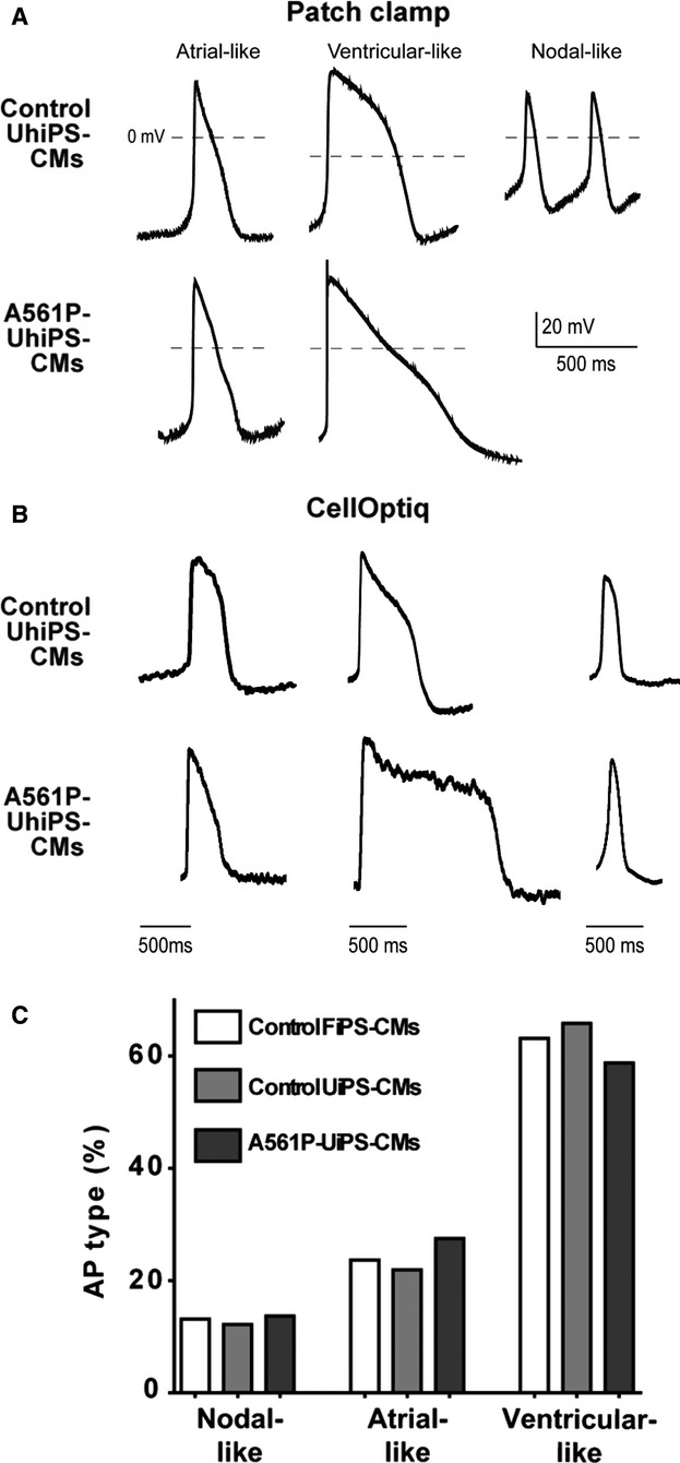
Figure 2.

Human UhiPS cells differentiated into electrically functional cardiomyocytes. Representative traces of spontaneous nodal-, atrial- and ventricular-like action potential recordings using (A) patch-clamp or (B) optical dye (CellOptiq) in control UhiPS-CMs and A561P-UhiPS CMs (no nodal-like AP was obtained in A561P-UhiPS-CMs using patch-clamp technique). C, Distribution of the 3 types of action potentials obtained by optical measurement. Regardless of the hiPS cell type, the majority of the action potentials were ventricular-like. Control FhiPS-CMs, n=38; control UhiPS-CMs, n=41; A561P-UhiPS CMs, n=51. AP indicates action potential; CM, cardiomyocytes; FhiPS, foreskin fibroblast–derived human induced pluripotent stem cells; UhiPS, urine-derived human induced pluripotent stem cells.
