Figure 1.

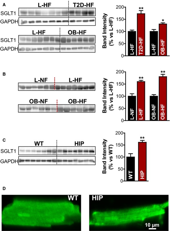
Increased SGLT1 protein expression in hearts from humans and rats with T2D. A, Western blots with an anti-SGLT1 antibody in homogenates of failing hearts from patients with T2D (T2D-HF group; 4 hearts) or obese (OB-HF group; 6 hearts) vs lean participants (L-HF group; 7 hearts). GAPDH was used as loading control, and experiments were repeated 4 times. Bar graph in the right panel shows the relative band intensity. B, SGLT1 expression in homogenates from failing vs nonfailing human hearts from lean (top) and obese (bottom) participants. C, Western blots with an anti-SGLT1 antibody in diabetic HIP vs WT heart homogenates. D, Representative immunofluorescence images of rat (WT and HIP) myocytes labeled with an anti-SGLT1 antibody. In both cases, SGLT1 is localized at the T-tubules. HF indicates heart failure; HIP, model of late-onset T2D; L, lean; NF, nonfailing heart; OB, obese; SGLT, Na+-glucose cotransporter; T2D, type 2 diabetes; WT, wild-type. *P<0.05, **P<0.01.
