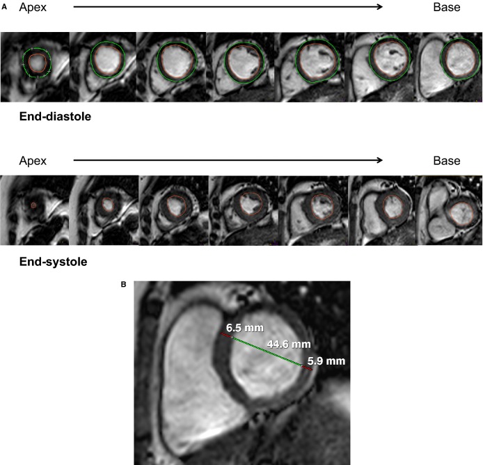
Figure 1.
Measurement of (A) left ventricular (LV) end-diastolic volume (top panel), end-systolic volume (bottom panel), and mass using manual tracing of endocardial (red) and epicardial (green) borders at end-diastole and tracing of endocardial border at end-systole and (B) LV wall thicknesses (red) and diameter (green) measurements taken at end-diastole from the short-axis slice immediately basal to the papillary muscles.

