
Figure 10.
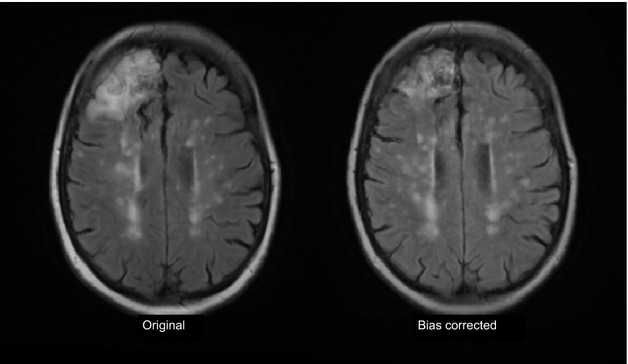
Effect of incautious use of bias field correction on cortical infarct and WMH. The left image shows the original MR FLAIR image with a right frontal cortical infarct (arrow) and numerous WMH. Following bias field correction (left) the frontal infarct appears much smaller, some of the WMH have become more (eg, posterior areas) and some now appear split into smaller components (mid centrum semiovale) as the bias field smoothing software tries to “even out” the distribution of signal intensities across the image. FLAIR indicates fluid attenuation inversion recovery; MR, magnetic resonance; WMH, white matter hyperintensities.