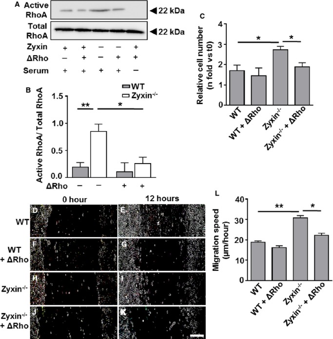
Figure 16.

Inhibition of RhoA activity reduces the proliferation and migration of Zyxin−/− VSMCs. A, Western blot of active RhoA immunoprecipitated from total cell lysates by Rhotekin Rho binding domain (RBD) beads 12 hours after exposure of the VSMCs to a Rho inhibitor (denoted by ΔRho) or left untreated. Serum-starved WT VSMCs were used as a negative control for Rho activation. B, Quantification of active RhoA: total RhoA ratio as compared to WT VSMCs. *P<0.05, **P<0.01 as indicated, n=4 for each experimental group. Total RhoA remained unchanged in WT and zyxin-deficient VSMCs. C, Statistical summary of proliferation of VSMCs, monitored by counting the number of cell nuclei stained with DAPI at 0 hour (t0) and after 24 hours (t24). The Rho inhibitor was added at 0 hour and again after 12 hours along with fresh medium. *P<0.05 as indicated, n=4 for each group. D through K, Representative images of the migration of WT and zyxin−/− VSMCs into a 2-dimensional scratch after 12 hours following treatment with the Rho inhibitor. Scale bar represents 500 μm. The sharpness and contrast of the images were adjusted to the same extent to clearly represent the migration front. L, Quantitative analysis of migration speed of the cells. The distance traveled by the cell front was divided by the time period to get the 2-dimensional migration speed. *P<0.05, **P<0.01 as indicated, n=4 for each group. DAPI indicates 4′,6-diamidino-2-phenylindole; VSMCs, vascular smooth muscle cells; WT, wild type.
