
Figure 1.
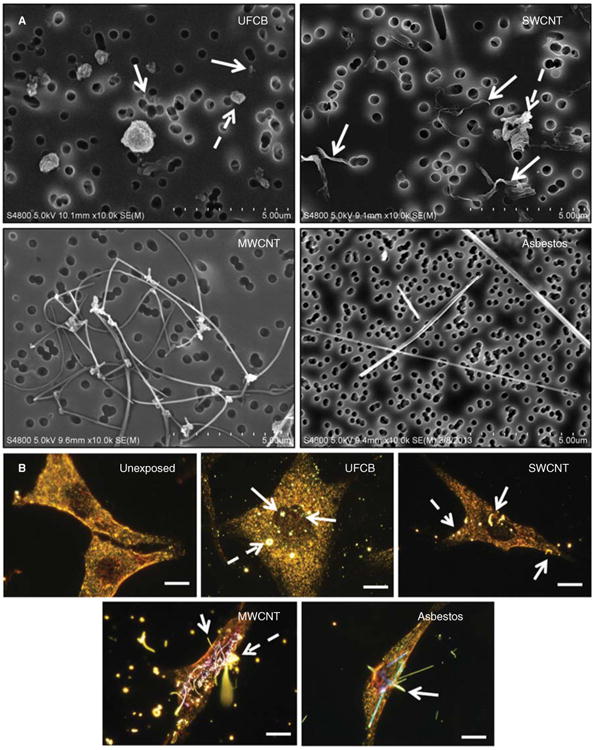
Visual characterisation of dispersed UFCB, SWCNTs, MWCNTs and crocidolite asbestos particle morphology and cellular fate in exposed human SAECs. A) Scanning electron microscope micrographs of dispersed particles. Dispersed UFCB (top left) and SWCNTs (top right) exhibited both nano-sized particles (white arrows) and micron-sized agglomerates (dashed arrow). MWCNTs (lower left) exhibited either single fibre or loosely tangled agglomerates while asbestos (lower right) existed as needle-like fibres. B) Hyperspectral dark-field microscopy micrographs of 24-h exposed SAEC to each dispersed particle. Cells were fixed and toluidine stained for contrast. Particles possess an intense white halo while stained cytoplasm and nucleus appears yellow and blue/purple, respectively. Nano-sized particles were co-localised to both cytoplasm and nucleus (solid while arrow) while micron-sized agglomerates were associated with cytoplasm or acellular (dashed white arrows). SWCNTs, MWCNTs and asbestos fibres were observed to penetrate and co-localise with both cytoplasm and nucleus. Bar = 10 μm.