
Figure 2.
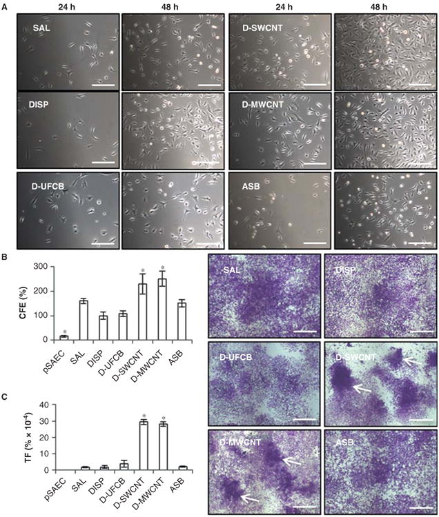
Altered proliferation and morphological transformation in SAECs subchronically exposed (6 months) to dispersed SWCNTs, dispersed MWCNTs or crocidolite asbestos. A) D-SWCNT and D-MWCNT cells showed greater density of cells at 48 h post-seeding than all other treatments. Bar = 500 μm. B) D-SWCNT and D-MWCNT cells possessed significantly greater CFE than DISP control and all other treatments while parental SAECs (pSAEC) exhibited low efficiency. C) D-SWCNT and D-MWCNT cells exhibited higher TF than all other treatments based on Type III foci counts (left). Type III foci (right; white arrows) exhibited deep basophilic staining, multi-layered cell growth and invasive growth into monolayer. All other groups demonstrated Type I foci with SAL and ASB cells occasionally displaying Type II foci. Bar = 1 mm.