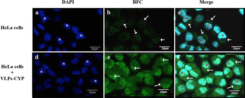
Fig. 3.

Cytochrome P450 activity assay in human cervix carcinoma cell line (HeLa). Staining with DAPI show nuclei of HeLa cells labeled as “n”, panels a and d. Endogenous CYP activity over BFC reagent was visualized in HeLa cells as observed in panel b. CYP activity of transfected VLPs-CYP nanoparticles in HeLa cells is shown in panel e. Overlay of DAPI and BFC localize the CYP activity in the cytoplasm of HeLa cells (white arrows), panels c and f. Scale bar represents 20 μm. Cells were visualized with a ×63 (DIC), 1.4 N.A. planapochromatic oil immersion objective
