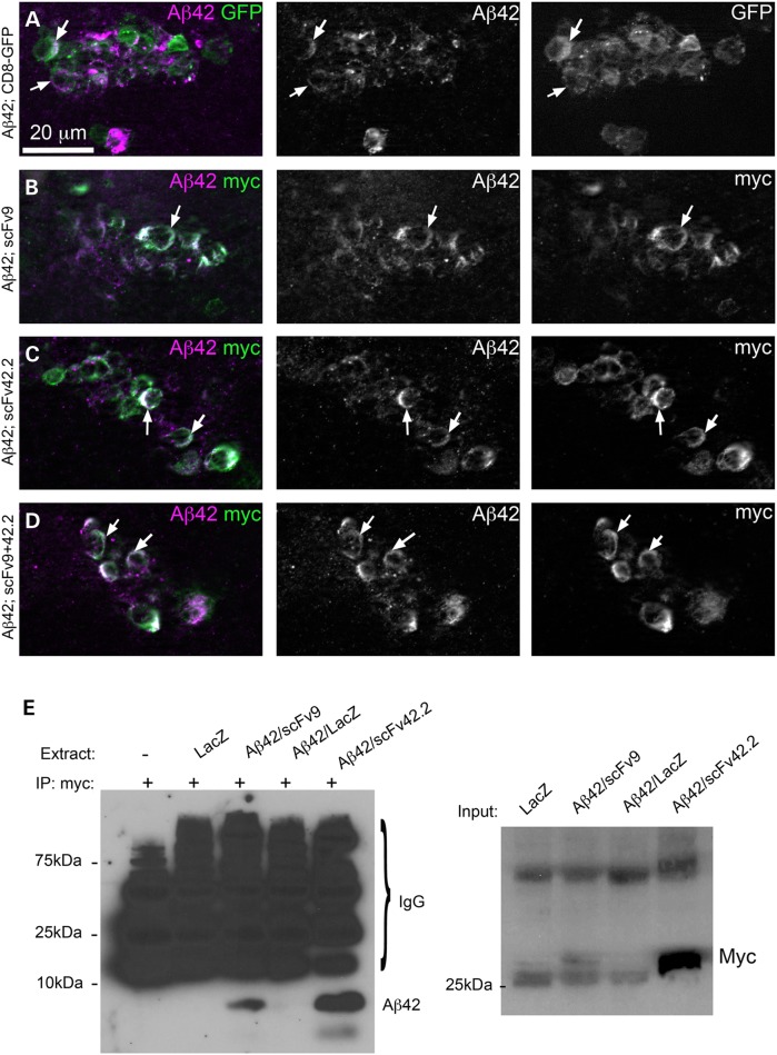
Figure 4.
Anti-Aß scFvs co-localize with Aß42 in brain neurons. (A–D) Subcellular distribution of Aß42 and anti-Aß scFvs in brain neurons. The transgenes are expressed under the control of OK107-Gal4 and localized in a small cluster of local interneurons of the antennal lobe. (A) Co-expression of Aß42 (magenta) and CD8-GFP (green) shows co-distribution in the membrane (arrows) and punctate accumulation of Aß42 in the Golgi. (B) Co-expression of Aß42 and scFv9 (myc epitope, green) results in co-localization in the membrane without changes in Aß42 distribution. (C) Co-expression of Aß42 and scFv42.2 (myc epitope, green) shows co-localization in the membrane and no changes in Aß42 distribution. (D) Co-expression of Aß42 together with scFv9 and scFv42.2 results in no evident changes in Aß42 distribution. (E) Aß42 and scFvs co-immunoprecipitate in Drosophila brain neurons. Using beads coated with the anti-myc antibody to capture the scFvs, we detected Aß42 immunoreactivity in samples co-expressing Aß42 and scFv9 or scFv42.2. Flies co-expressing Aß42 and scFv42.2 produced higher Aß42 immunoreactivity, consistent with its higher expression levels. Three negative controls produced non-specific signal from antibody fragments released from the column. On the right, we show the input for the co-IP immunoblotted against anti-myc. As expected, the levels of scFv42.2 are several times higher than those of scFv9.

