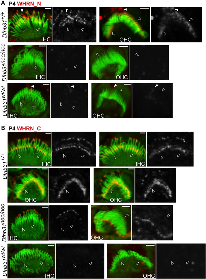
Figure 2.
Whirlin protein localization in developing wild-type, Dfnb31neo/neo and Dfnb31wi/wi cochlear hair cells. (A) Immunostaining using rabbit WHRN_N antibody shows signals at the tip and base of IHC stereocilia and only at the base of OHC stereocilia in P4 wild type mice. The same experimental procedure detected no signals in P4 Dfnb31neo/neo and Dfnb31wi/wi cochlear stereocilia. Note that rabbit WHRN-N antibody also detected signals along the kinocilium in P4 wild-type and Dfnb31wi/wi cochlear hair cells (white filled arrows). (B) Immunofluorescence using rabbit WHRN_C antibody shows signals at the tip and base of both IHC and OHC stereocilia in P4 wild-type mice. In P4 Dfnb31neo/neo IHCs and OHCs, immunoreactivity of rabbit WHRN_C antibody was found at the stereociliary tip. No immunoreactivity of WHRN_C antibody was detected in P4 Dfnb31wi/wi cochlear stereociliary bundles. Empty arrows point to stereociliary bases. The red signals of whirlin proteins are shown in grayscale on the right of each overlay panel with the matched position of arrows. Red signals outside stereociliary bundles are non-specific. Scale bars, 1 µm.

