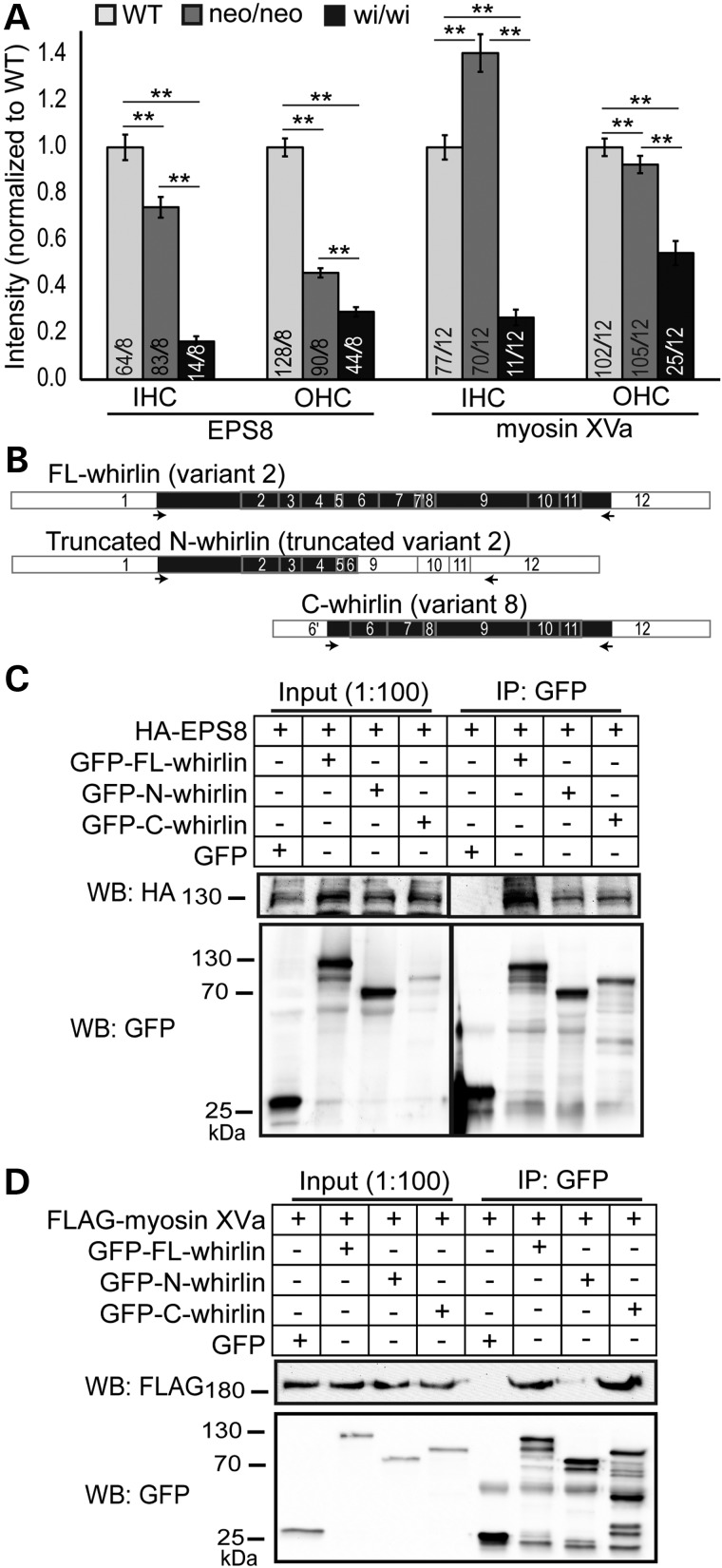
Figure 6.
EPS8 and myosin XVa expressions in Dfnb31neo/neo and Dfnb31wi/wi cochlear hair cells and their interactions with whirlin fragments. (A) Quantification of EPS8 and myosin XVa expressions in wild-type (WT), Dfnb31neo/neo (neo/neo) and Dfnb31wi/wi (wi/wi) mice. EPS8 expression was reduced at the IHC and OHC stereociliary tip of both Dfnb31neo/neo and Dfnb31wi/wi mice. EPS8 expression was less in Dfnb31wi/wi hair cells than in Dfnb31neo/neo hair cells. Myosin XVa expression was increased in IHC stereocilia but decreased in OHC stereocilia of Dfnb31neo/neo mice, while myosin XVa expression was reduced in both IHC and OHC stereocilia of Dfnb31wi/wi mice. Numbers of cells and mice analyzed are shown in the bottom of each bar before and after slashes, respectively. Error bars represent standard error of the mean. Student's t-tests (two-tail) were performed. **P < 0.01. (B) Schematic diagram of whirlin protein fragments used in the coimmunoprecipitation experiments shown in (C) and (D). Arabic numerals are exon numbers. White and black colors indicate untranslated and protein coding regions, respectively. cDNA fragments flanked by arrows were cloned into the GFP-tagged expression vectors. (C) HA-tagged EPS8 protein was able to be coimmunoprecipitated with GFP-tagged FL-whirlin, truncated N-whirlin and C-whirlin. (D) FLAG-tagged myosin XVa tail fragment was able to be coimmunoprecipitated with GFP-tagged FL-whirlin and C-whirlin but not the truncated N-whirlin. GFP protein from the empty pEGFP-C vector was used as a negative control. The anti-GFP blots demonstrate the success of immunoprecipitations and the amounts of GFP-tagged proteins pulled down in the experiments.

