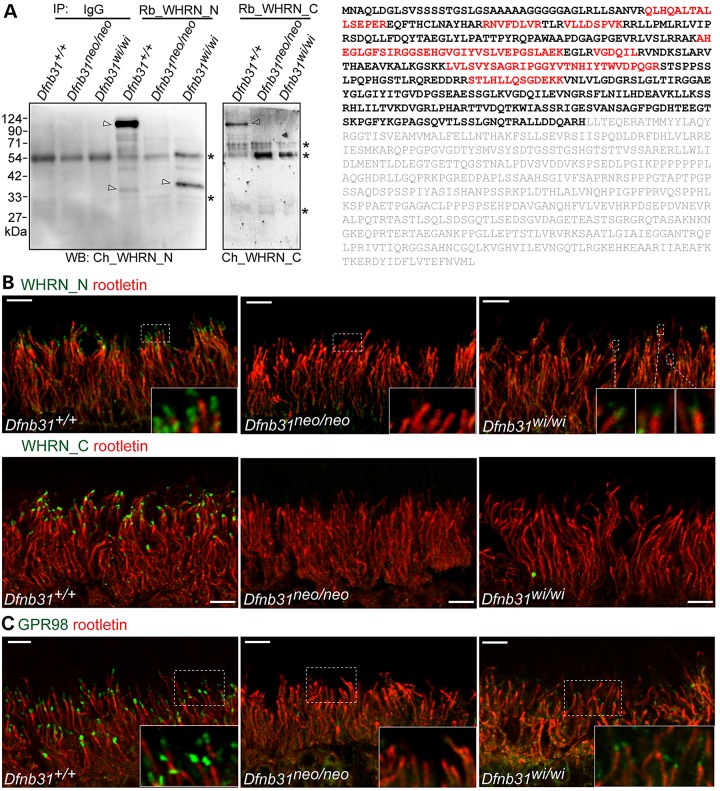
Figure 9.
Whirlin protein expression, localization and function in Dfnb31neo/neo and Dfnb31wi/wi photoreceptors. (A) Left, immunoblotting of whirlin immunoprecipitates from adult wild-type, Dfnb31neo/neo and Dfnb31wi/wi retinas. Arrows point to whirlin-specific bands, and asterisks label the antibody or non-specific bands. IP, immunoprecipitation; WB, immunoblotting; IgG, rabbit immunoglobulin, a negative control; Rb_WHRN_N and Rb_WHRN_C, rabbit antibodies against whirlin N- and C-terminal regions, respectively (Fig. 1A); Ch_WHRN_N and Ch_WHRN_C, chicken antibodies against whirlin N- and C-terminal regions, respectively (Fig. 1A). Right, peptides identified by mass spectrometry are labeled in red in the amino acid sequence of whirlin isoform 2 (NP_001008791). Amino acids labeled in gray are after the Dfnb31wi mutation. (B) Immunostaining of wild-type, Dfnb31neo/neo and Dfnb31wi/wi retinas using rabbit WHRN_N (upper row) and WHRN_C (lower row) antibodies. Residual whirlin signals (green) were detected above the ciliary rootlet (rootletin, red) at the periciliary membrane complex in Dfnb31wi/wi but not Dfnb31neo/neo photoreceptors using the WHRN_N antibody, while no whirlin signals were found in Dfnb31neo/neo or Dfnb31wi/wi photoreceptors using the WHRN_C antibody. Insets are the amplified view of regions framed by white dashed lines. (C) Residual GPR98 signals (green) were detected above the ciliary rootlet (rootletin, red) at the periciliary membrane complex in Dfnb31wi/wi but not Dfnb31neo/neo photoreceptors. Insets are the enlarged view of GPR98 signals in white boxes. Scale bars, 5 µm.

