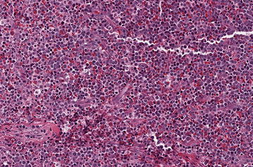
Fig. 1.

Hematoxylin and eosin stained slide of left neck biopsy. A diffuse proliferation of intermediate to large mononuclear cells with round to irregular nuclei, dispersed chromatin, distinct nucleoli, and small amounts of cytoplasm, consistent with blast forms are seen. Admixed are plasma cells, small lymphocytes, and additional myeloid elements, including abundant eosinophilic forms
