Fig. 3.

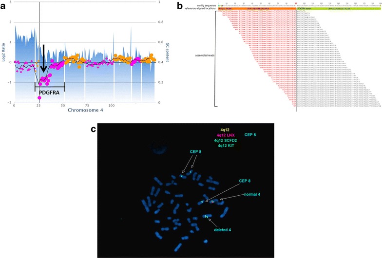
Molecular diagnostics of the patient’s tumor. a Copy number assessment of chromosome 4 shows a one copy loss of the 5′ end of PDGFRA. b Translocation analysis shows the discordant reads map to intron 10 of FIP1L1 and exon 12 of PDFGRA. c Metaphase FISH analysis shows one normal copy of chromosome 4, which retains all 3 FISH probes on 4q (green SCFD2 that is centromeric to FIP1L1, orange LNX that is located between FIPL1 and PDGFRA, and blue KIT that is telomeric to PDGFRA). The other copy of chromosome 4 shows an isolated deletion of LNX with retention of the flanking SCFD2 and KIT probes, indicative of a FIP1L1-PDGFRA rearrangement. Trisomy 8 is also present in these cells, as evidenced by 3 CEP 8 probe signals
