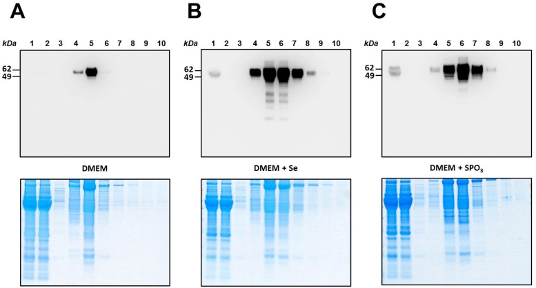
Fig 3. Isolation of SelP from culture media of HepG2 cells.
Medium samples from HepG2 cells were chromatographed on HisPur resin. Fractions were analyzed by SDS-PAGE, the gels stained with Coomassie blue (Lower panels) and subjected to Western blotting with anti-SelP antibodies (Upper panels). Lane 1, initial sample, lane 2, flow-though fraction, lane 3, buffer wash, lane 4–10, elution with a gradient of imidazole from 0 to 200 mM in loading buffer. (A) HepG2 cells grown on DMEM medium only (control). (B) HepG2 cells grown on DMEM supplemented with 100 nM Se. (C) HepG2 cells grown on DMEM supplemented with 1 mM thiophosphate (SPO3). Protein molecular weights markers in kDa are shown on the left. Experimental details are given in Materials and Methods.

