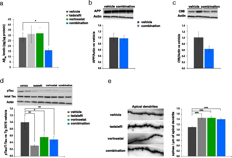
Fig. 4.

Chronic treatment (4 weeks) with the combination of vorinostat and tadalafil reversed the AD histopathological markers in aged Tg2576 mice. a Aβ42 levels determined by ELISA in parieto-temporal cortex of Tg2576 mice treated with vehicle, vorinostat, tadalafil, or combination (vorinostat and tadalafil) (n = 8–10 per group) (*p ≤ 0.05). In this and all subsequent figures, results are expressed as mean ± SEM. Effects of chronic administration of vorinostat and tadalafil on full-length APP (b) or C-terminal fragment C99 (c) levels, normalized to actin, in SDS parieto-cortical extracts. The histogram shows the quantification of the immunochemically reactive bands in the Western blot (representative bands are shown). Data are expressed as the fold change versus Tg2576 mice receiving vehicle (n = 8–10 in each group). d Representative Western blot bands using the antibody AT8 (pTau), normalized to total tau (T46) of cortical tissues are shown. The histograms represent the quantification of the immunochemically reactive bands in the Western blot. Data are expressed as the fold change versus Tg2576 mice receiving vehicle (n = 8–10 in each group) (*p ≤ 0.05; **p ≤ 0.01). e Representative Golgi staining images of apical dendrites on CA1 hippocampal pyramidal neurons. Scale bar, 10 μm. The histograms represent the quantification of spine density of apical dendrites on hippocampal CA1 pyramidal neurons from Tg2576 mice treated with vehicle, vorinostat, tadalafil or combination (vorinostat and tadalafil) (n = 34–36 neurons from three to four animals per group) (***p ≤ 0.001)
