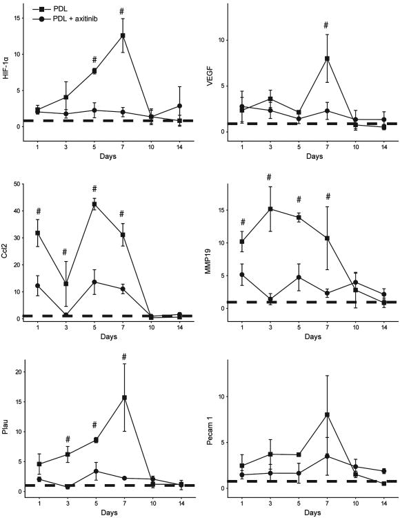
Fig 2.
Topical application of axitinib significantly suppressed PDL-induced mRNA levels of angiogenic factors. The relative mRNA levels of hypoxia-inducible factor (HIF)-1α, vascular endothelial growth factor (VEGF), chemokine (C-C motif) ligand 2 (Ccl2), matrix metalloproteinase (MMP)19, plasminogen activator urokinase (Plau) and platelet endothelial cell adhesion molecule (Pecam) 1 were plotted against days post-PDL exposure. The data are presented as mean ± SD and show the fold changes of mRNA levels of target genes (y-axes) compared with the normal controls (dashed lines). #P < 0.05 in the PDL only group compared with the PDL + axitinib group (paired t-test).

