
Fig 3.
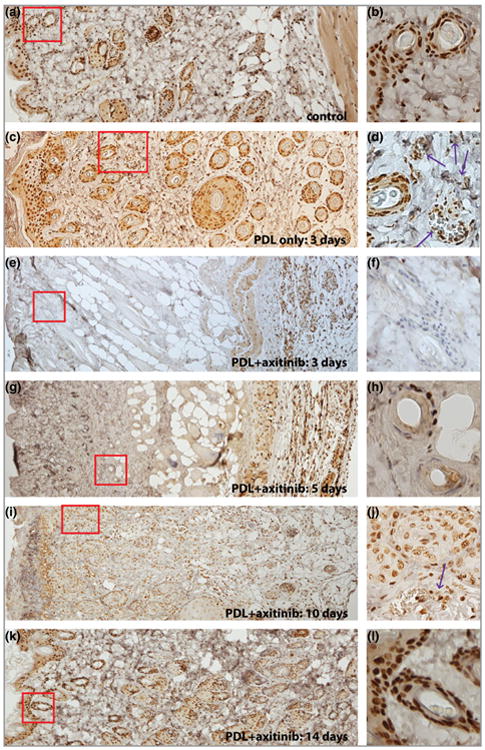
Topical application of axitinib significantly blocked PDL-induced activation of extracellular regulated kinase (ERK). (a, b) Epidermis and hair follicles showed strong basal activity of ERK. (c, d) The activity of ERK was elevated in the photocoagulated blood vessels (indicated by the arrows) in the dermis 3 days post-PDL exposure. (e–h) The activity of ERK in the epidermis and dermis, but not in muscular layers, was blocked by administration of 0.5% topical axitinib for (e, f) 3 or (g, h) 5 days. (i, j) The activity of ERK was partially restored in the dermis, but not the epidermis, 10 days post-PFL exposure. Note the weaker intensity of phosphor-ERK (pERK) immunoreactive signals of hair follicles in (j) compared with the basal activity of ERK in (b). (k, l). The pERK expression pattern in both the epidermis and dermis was back to normal 14 days post-PDL exposure. The images in the right-hand column are higher magnifications of the boxed areas in the corresponding left-hand panels. Red boxes highlight epidermis/dermis or dermis only.