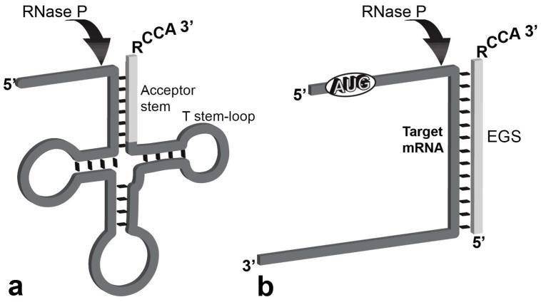
Fig. 1.
Cleavage of a pre-tRNA and an mRNA. (A) The arrow shows the site of action of RNase P on a pre-tRNA. The clear segment is the acceptor stem. (B) Complex between a target mRNA and an EGS that can be recognized as substrate by bacterial RNase P. In this example, the ATG sequence has been added to represent the possibility of using an mRNA as target. The RCCA sequence mimics the 3′-end of the pre-tRNA and facilitates interaction with RNase P. Redrawn from Ref. 4.

