Figure 3.
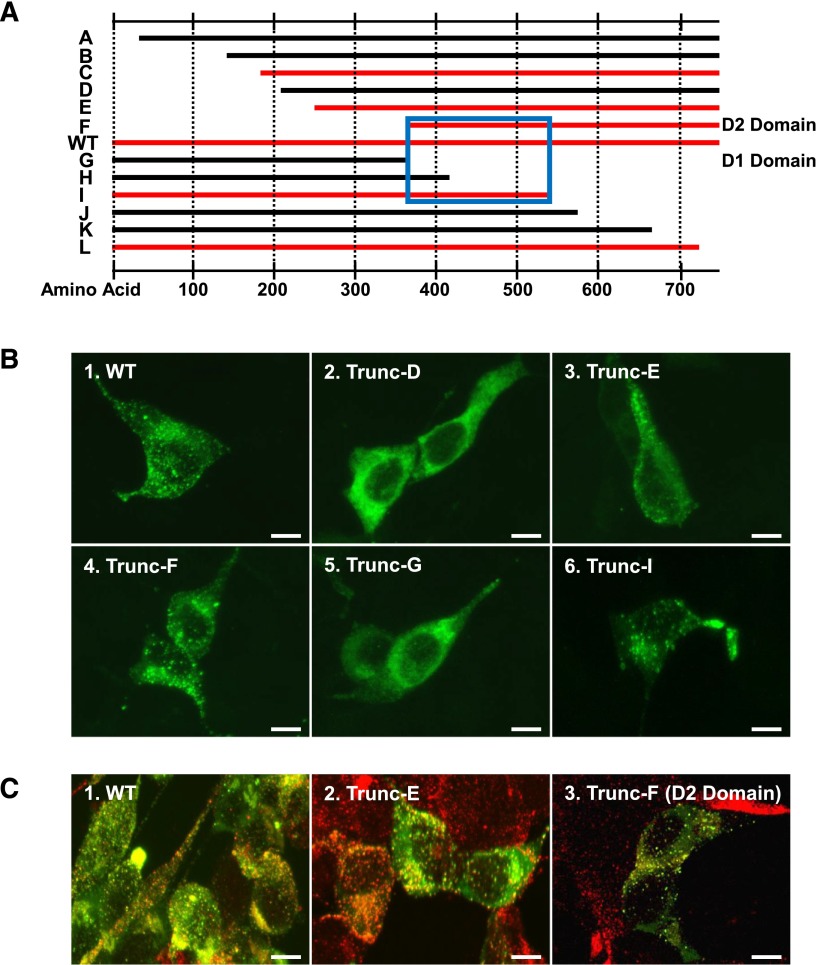
Location of VWF “storage signal.” (A) Schematic representation of N-terminal and C-terminal truncations of VWFpp. Each construct contains the VWF signal peptide followed directly by the VWFpp sequence depicted. WT VWFpp is located in the middle. VWFpp truncations that were observed to traffic to storage granules are shown in red, whereas those truncations that appeared to be ER-localized are shown in black. The minimum area of sequence overlap between truncations that sorted to storage granules is highlighted in the blue box. (B) AtT-20 cells were transiently transfected with VWFpp truncation constructs to determine the region of VWFpp containing the VWF “storage signal.” After 72 hours, cells were fixed, permeabilized, stained with monoclonal antibodies to VWFpp, and detected with Alexa Fluor-488–conjugated goat anti-mouse immunoglobulin (Ig)G (green) by confocal microscopy. Expression of VWFpp truncations E, F, and I (panels 3, 4, and 6) resulted in granular storage of VWFpp similar to WT (panel 1), whereas truncations D and G appeared to be ER-localized (panels 2 and 5). (C) AtT-20 cells were transiently transfected with VWFpp truncations to address co-localization with endogenous ACTH-containing granules. After 72 hours, cells were fixed, permeabilized, dual stained with monoclonal antibodies to VWFpp and a polyclonal antibody to ACTH, and detected with Alexa Fluor-488–conjugated goat anti-mouse IgG (VWFpp/green) and Alexa Fluor-594–conjugated goat anti-rabbit IgG (ACTH/red) by confocal microscopy. Colocalization of VWFpp and ACTH is shown in yellow. Truncations E and F (panels 2 and 3) co-localized with endogenous ACTH-containing granules similar to WT VWFpp (panel 1). Total magnification, ×1500. Bar, 10 μm. Collectively, these results demonstrate that VWFpp contains a “storage signal” that appears to be localized in the D2 domain of VWFpp.

