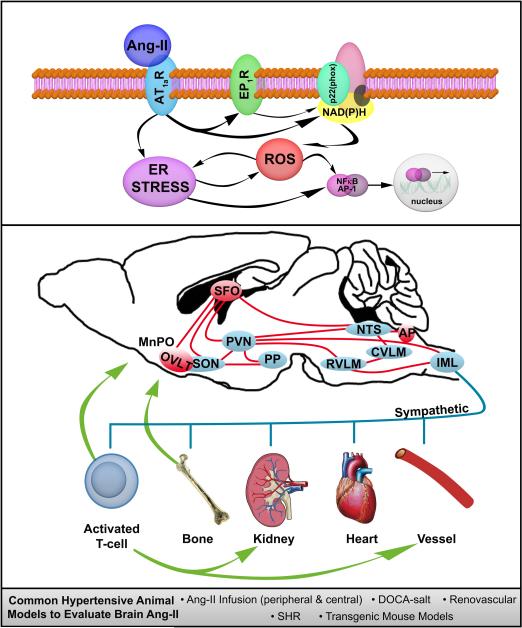
FIGURE.
Simplified schematic illustrating the signaling pathways (top box), as well as neural networks and sympathetic nervous system influenced physiological outputs (middle box) involved in the development of hypertension due to peripherally- or locally-generated Angiotensin-II (Ang-II) action in the brain. Common animal models used to evaluate Ang-II, the brain and hypertension are also highlighted (bottom box). See text for additional details. AP, area postrema; AP-1, activator protein-1; AT1aR, Angiotensin type 1a receptor; CVLM, caudal ventral lateral medulla; DOCA, deoxycorticosterone acetate; ER, endoplasmic reticulum; EP1R, prostaglandin E receptor 1; IML; intermediolateral nucleus; MnPO, median preoptic nucleus; NAD(P)H, nicotinamide adenine dinucleotide phosphate; NTS, nucleus tractus solitarii; NFκB, nuclear factor κB; OVLT, organum vasculosum lamina terminalis; PP, posterior pituitary; PVN, paraventricular nucleus of the hypothalamus; ROS, reactive oxygen species; RVLM, rostral ventral lateral medulla; SHR, spontaneously hypertensive rat; SFO, subfornical organ; SON, supraoptic nucleus.

