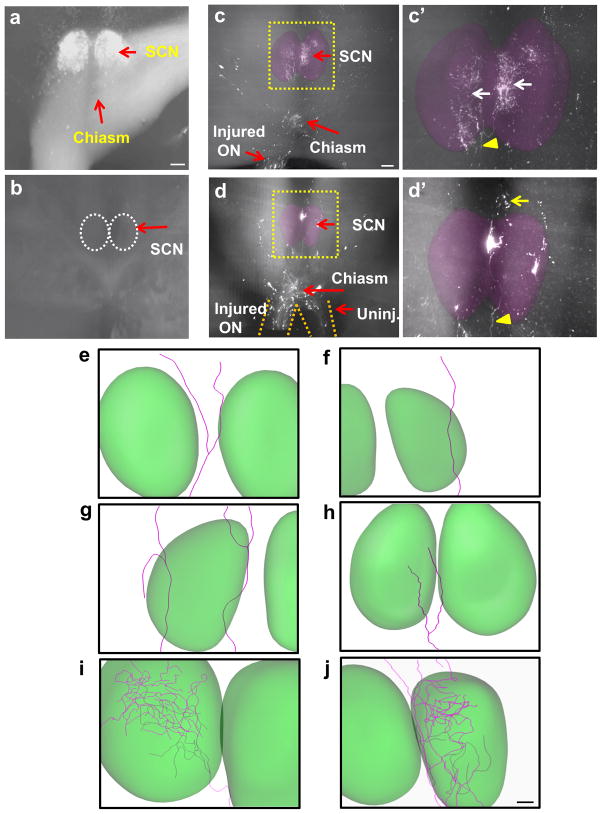
Figure 5. Ultramicroscopic visualization of axonal projections in mouse visual target.
(a) Optic chiasm, optic tract and SCN shown by CTB labeling in a cleared, uninjured brain. (b) Bottom view of a brain containing the SCN (i.e. dotted circles) imaged from a brain of an injured control mouse subjected to AAV-EGFP injection. None of the control animals had regenerating axons in the brain. (c) and (d) Bottom view of brains of two different PCC-treated animals showing regenerated axons in the chiasm and SCN. Yellow arrowhead indicates a regenerating axon extending into the SCN. Yellow arrow indicates distal end of the same axon showing continued growth beyond the SCN. White arrows in d indicate extensive arborization. (e–j), reconstructions of single axons showing various growth patterns. In (e), an axon bypasses the target. In (f) and (g), axons traverse the target. In (h), axon terminals are found within the SCN. In (i) and (j), axons enter the SCN and form complex arborization. ON, optic nerve; Uninj, uninjured; SCN, suprachiasmatic nucleus. Scale bars, 100 μm.

