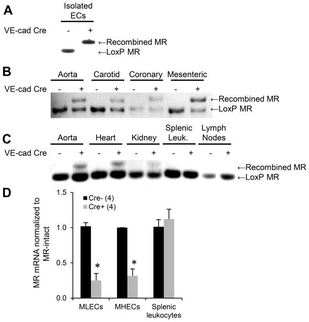
Figure 1. A mouse model with MR deleted specifically from EC and not leukocytes.
(A–C) MR genomic DNA was amplified with primers specific for the LoxP MR or recombined MR. (A) The MR gene is completely recombined in primary cultured mouse ECs only from Cre+ mice. (B) MR recombination occurs in Cre+ aorta, carotid, coronary, and mesenteric vessels. (C) MR recombination occurs only in EC-containing tissues in VE-cad-Cre+ mice and not in lymph nodes or splenic leukocytes (Leuk.) (D) MR mRNA is reduced in primary ECs cultured from mouse lungs (mouse lung endothelial cells, MLECs) and hearts (MHECs) but not in leukocytes from EC-MR-KO mice. Number of animals per group is indicated in parentheses. *p<0.05 versus MR-intact.

