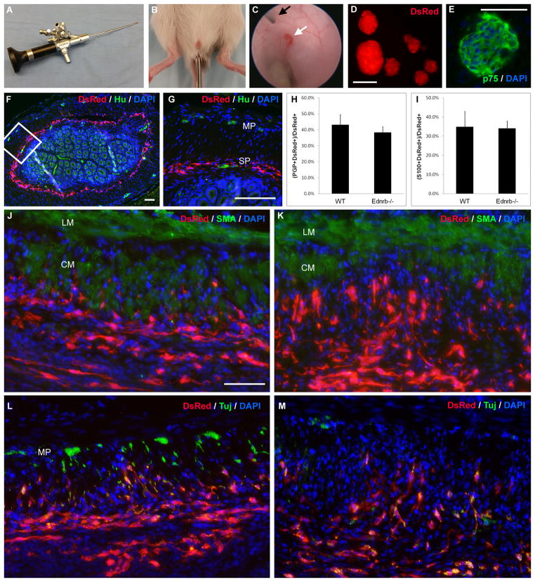
Figure 1. Enteric neuronal stem cells can be delivered by colonoscopy into the gut wall.
Colonoscopy is performed using a 4.5 Fr cystoureteroscope (A). The endoscope is introduced into the anus of WT or Ednrb−/− mice and advanced 2 cm (B). A 30-gauge Hamilton needle (C, black arrow) is passed through the working channel of the endoscope and 50 μL of a 1,000 cells μL−1 suspension is injected into the colon wall (C, white arrow). Enteric neurospheres generated from Actb-DsRed mice ubiquitously express DsRed (D) and also stain for neural progenitor marker, p75 (E). Immediately following endoscopic delivery of cells, there is circumferential spreading of DsRed-positive cells in the region of the submucosal plexus of the distal colon (F, inset magnified in G). Endogenous enteric neurons are labeled by neuronal marker, Hu, in a WT recipient (G). One week after injection, there is no significant difference in neuronal (H) or glial density (I) among transplanted cells in WT and Ednrb−/− recipients. Distribution of transplanted cells is similar in WT (J) and Ednrb−/− (K) recipients 1 week after transplantation. Fibers co-expressing neuronal marker, Tuj, extend into the muscularis propria both in WT (L) and Ednrb−/− (M) recipients. Endogenous myenteric ganglia are seen in WT colon (L). Scale bar is 100 μm. Scale bar is the same for J–M.
CM, circular muscle; LM, longitudinal muscle; MP, myenteric plexus; SP, submucosal plexus

