FIG 3 .
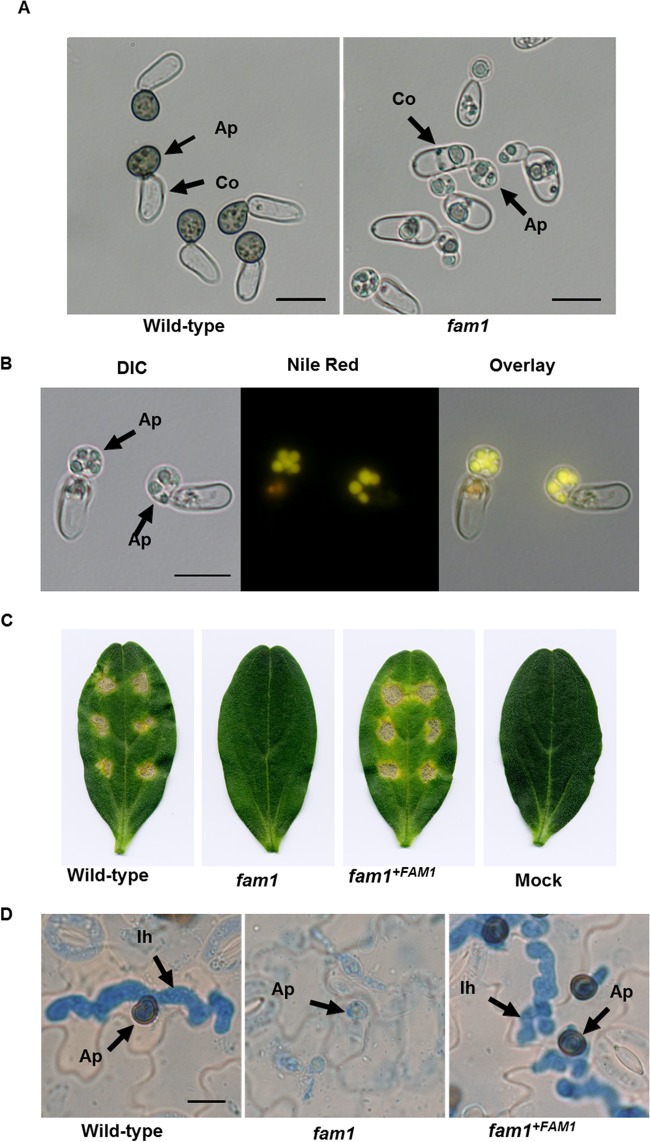
Appressorium development and pathogenicity of the fam1 mutant. (A) Conidia of the wild-type strain or fam1 mutant were incubated on glass slides for 24 h and observed by light microscopy. Ap, Appressoria; Co, conidia. Bars, 10 µm. (B) Distribution of lipid droplets in appressoria of the fam1 mutant. After the cells were stained with Nile red, they were viewed with differential interference contrast (DIC) microscopy, epifluorescence microscopy, and as an overlay. Bar, 10 µm. (C) The fam1 mutant showed attenuated pathogenicity on cucumber cotyledons. Cotyledons were inoculated with spores of the wild type, fam1 mutant, complemented strain, or distilled water (mock). Symptoms were observed after 6 days. (D) Histology of infection. Spores of the wild-type, fam1 mutant, and complemented strain were inoculated on cucumber cotyledons, and tissues were examined by light microscopy after 72 h. Ap, appressoria; Ih, infection hyphae. Bar, 10 µm.

