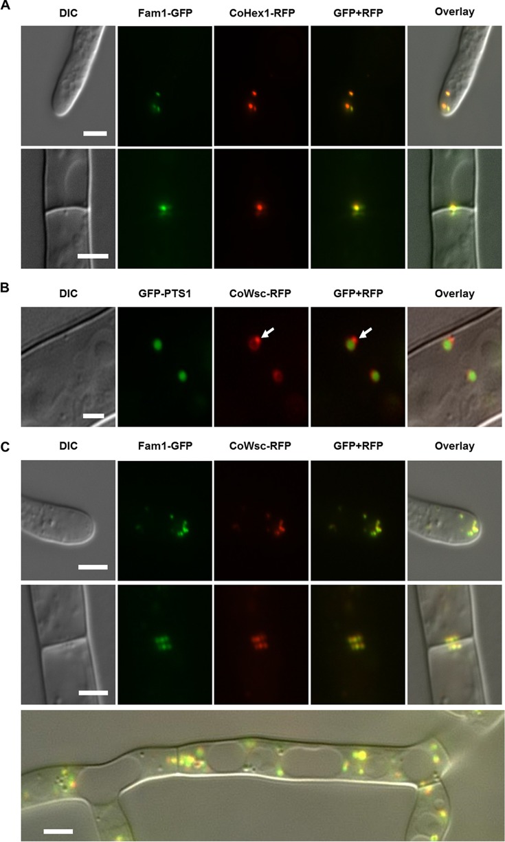
FIG 5 .
Fam1 is localized in Woronin bodies. Images show vegetative hyphae viewed by differential interference contrast (DIC) microscopy, epifluorescence of GFP or RFP, merged GFP and RFP channels, and an overlay of all channels. (A) In hyphae coexpressing Fam1-GFP and CoHex1-RFP, Fam1-GFP colocalized with Woronin body (WB) matrix protein CoHex1 on WBs at hyphal tips (top panels) and septa (bottom panels). Bars, 4 µm. (B) In hyphae coexpressing GFP-PTS1 and CoWsc-RFP, the WB peripheral membrane protein CoWsc localized around the peroxisomal matrix (labeled by GFP-PTS1) and was enriched at sites where nascent WBs budded from mother peroxisomes (white arrows). Bar, 2 µm. (C) In hyphae coexpressing Fam1-GFP and CoWsc-RFP, both markers colocalized in WBs at hyphal tips (top panels), septa (middle panels), and in the cytoplasm of subapical cells (bottom panel, merge image). Bars, 2 µm.

