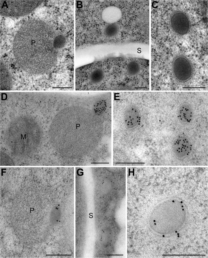
FIG 6 .
Immunoelectron microscopic localization of Fam1 in Woronin bodies. TEM images showing the ultrastructure of peroxisomes and Woronin bodies in vegetative hyphae of C. orbiculare (A to C) and the immunogold localization of Hex1 (D and E) and Fam1 (F to H). (A) Peroxisome (P) with nascent WB budding from it. (B) Mature WBs in the cytoplasm near a septum (S). (C) The single membrane of mature WBs is thicker than the peroxisome membrane and has a prominent bilayer structure. (D and E) Hex1 is localized in the dense matrix of nascent and mature WBs. (F to H) Fam1 is localized in the membranes of nascent WBs (F) and mature WBs (G and H) but is not detected on the peroxisome membrane. Bars, 200 nm.

