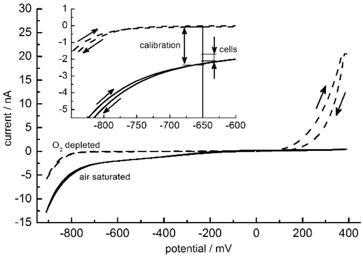
Figure 7.
Mean of 10 cyclic voltammograms of a 25-µm oxygen electrode in two-electrode configuration between −900 and 400 mV (step potential 10 mV, scan rate 10 mV/s) for air-saturated (solid lines) and oxygen-free media (dashed lines). Voltammetric scan directions are marked by arrows. Insert: zoom around −650 mV. Double arrows mark the current ranges swept during calibration of the electrode (−2.346 ± 0.038 nA (oxygen saturated); −0.073 ± 0.007 nA (oxygen depleted)) and during cell culture at −650 mV (Figure 8). Please note that the steep current increase for the oxygen-free medium above 100 mV is induced by the presence of 1% sodium sulphite.

