Fig. 3.

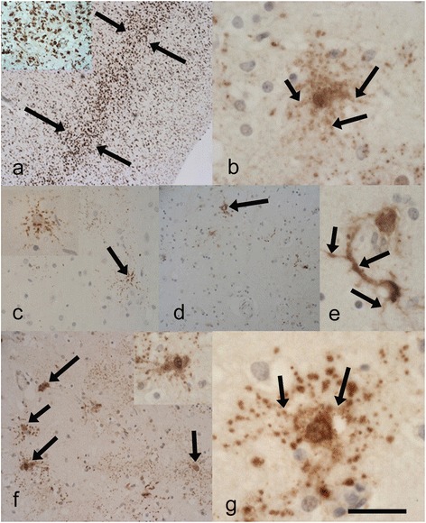
Case 6 (p.P525L and p.Y374X TARDBP mutation) (a) and inset illustrates marked microglial activity in the mid-deeper laminae (between arrows) of the motor cortex. Anti-CD68. b Showing the unusual neuronal staining for p62 in the motor cortex including processes (arrows). c-e revealing the sparse but unusual neuronal p62 immunopositivity in the hippocampal region (c) and (c) inset including occasional NCIs (arrow), and (d) the putamen revealing occasional NCIs (arrow). e Reveals one of the few p62 immunopositive neurons in the putamen, this is also apparently labelling the corresponding axon/dendrites (arrows) and appears different in pattern to the occasional FUS positive NCIs. P62 immunopositivity in the frontal neocortex (f) revealing dot-like positivity and neuronal inclusions (arrows and inset). g p62 immunopositive neuron in the temporal neocortex. These inclusions are negative for FUS and TDP-43 and appear to label the nuclear/ perinuclear region and processes (arrows) the latter indicating likely axonal/dendritic positivity. Anti-p62. Scale Bar (a)-300 μm, a-inset 100 μm (b)-50 μm, (c)-150 μm, (c) inset-80 μm, (d) 200 μm, (e)-50 μm, (f)-200 μm, (f) inset-80 μm, (g)-30 μm
