Figure 2.

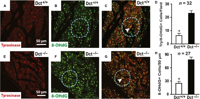
Dct reduces oxidative modifications in CMLCs and the surrounding myocardium. Representative images from wild-type (A–C) and Dct-null (E–G) atria costained with antibodies against tyrosinase (red) and 8-OHdG (green). (A) and (E) show antityrosinase staining, (B) and (F) show anti-8-OHdG staining, and (C) and (G) show merged images using triple filter settings. In (C) and (G), the arrowhead points to CMLCs (yellow) that costain for both tyrosinase (green) and 8-OHdG (red). In (B–C) and (F–G), the blue dashed circle denotes a 50-μm radius surrounding the costained CMLC. The scale bar in (A) applies to (A–C), and scale bar in (E) applies to (E–G). In (D) the bar graph compares the number of cells costained positive for 8-OHdG and tyrosinase per 20× field in Dct-null and wild-type atria. In (H) the bar graph compares the number of anti-8-OHdG stained nuclei within a 50-μm radius of Dct-null and wild-type CMLCs costained positive for tyrosinase and 8-OHdG. At least 27 fields were analyzed from three different hearts in each group. Error bars represent the standard deviation. *P < 0.05.
