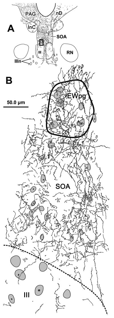
Figure 3.
The pattern of axonal labeling from a BDA injection of the cMRF in a section located near the rostral pole of the oculomotor nucleus (III) (A). The box indicates the region shown in B, which includes the preganglionic Edinger-Westphal nucleus (EWpg) and the portion of the supraoculomotor area (SOA) between EWpg and III. The axons show few branches, display primarily en passant boutonal enlargements, and densely populate this region. They cross the EWpg border freely, but tail off when crossing the border of III. Cresyl violet stained cells are indicated by shading.

