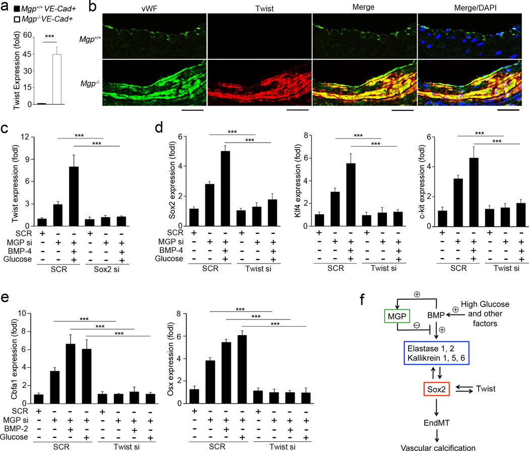
Figure 8. Mutual regulation of Sox2 and Twist1 in EndMTs in vascular calcification.
(a) Increase of Twist1 expression in VE-cadherin+CD45− presorted cells from Mgp−/− aortas, as shown by real-time PCR. ***p<0.001. (b) Co-localization of Twist1 with the EC marker vWF in Mgp−/− aortas, as shown by immunostaining. Scale bars: 100 µm. (c) MGP-depleted HAECs were transfected with siRNAs (si) to Sox2, and then treated with combinations of BMP-4 and glucose. Expression of Twist1 was examined by real-time PCR. ***p<0.001. (d) MGP-depleted HAECs were transfected with siRNAs (si) to Twist1, and then treated with combination of BMP-4 and glucose. Expression of Sox2, Klf4 and c-kit was examined by real-time PCR. ***p<0.001. (e) MGP-depleted HAECs were transfected with siRNAs (si) to Twist1, and then treated with BMP-2 or glucose for 4 days. Expression of Sox2, Klf4 and c-kit was examined by real-time PCR. ***p<0.001. (f) Schematic working model for the induction of serine proteases and Sox2 in EndMTs in vascular calcification.

