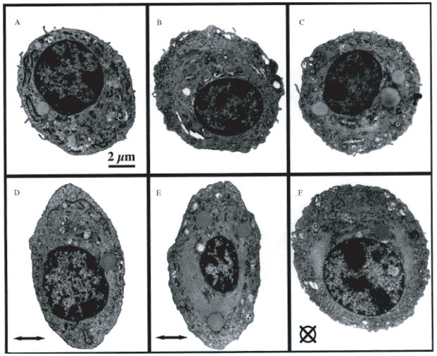
Figure 6.
Transmission electron micrographs of condrocytes embedded in agarose constructs held unstrained (A–C) or subjected to 20% compression (D–F). All micrographs were taken at the same magnification (Scale bar=2 μm). The direction of the applied strain is indicated by the horizontal arrow in (D) and (E) and a crossed circle in (F).
(Figure taken from Lee at al. J Biomechanics. 2000. 33: 81–85).

