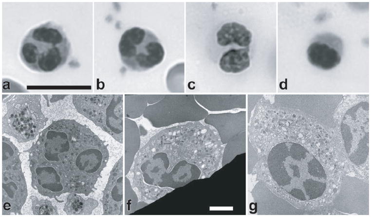
Figure 9.
Defects in nuclear morphology in neutrophils from blood smears of individuals with Pelger-Huet anomaly (PHA) labeled with Wright-Giemsa stain. (A) Normal neutrophil with lobulated nucleus. (B) Heterozygous PHA neutrophil with a bilobed nucleus, taken from the mother of the homozygous individual. (C) Heterozygous PHA neutrophil with a bilobed nucleus, taken from the father of the homozygote. (D) Homozygous PHA neutrophil showing an ovoid nucleus with chromatin clumping. Scale bar for light micrographs: 10 μm. (E–G) Transmission electron micrographs of normal human neutrophil nucleus with three apparent lobes and extensive peripheral heterochromatin (E), heterozygous PHA neutrophil with a bilobed nucleus taken from the father of the homozygote (F), and ovoid nucleus from a homozygous PHA granulocyte exhibiting extensive heterochromatin redistribution (G). Scale bar for electron micrographs: 1 μm
(Figure taken from Hoffmann et al. Chromosoma. 2007. 116: 227–235)

