
Figure 2.
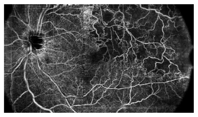
OCT angiography (composition map of three partially overlapping 8 × 8 mm scans) of a 57-year-old woman with branch retinal vein occlusion (BRVO) showing enlargement of foveal avascular zone and retinal nonperfused area (capillary drop-out) at the posterior pole and at the mid periphery with the development of collaterals.