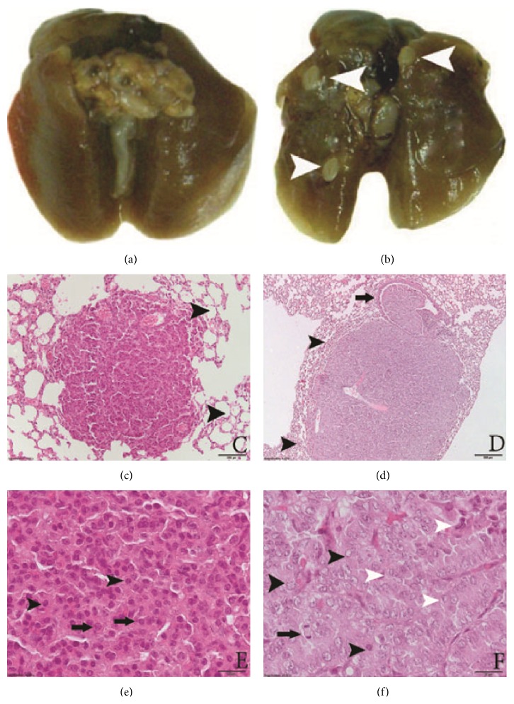
Figure 1.
Lung specimens. (a) Control. (b) Urethane-induced lung tumors (white arrowheads). (c) Adenoma, magnification ×4, H & E staining. There are no signs of pressure to surrounding tissues; alveoli are well preserved (black arrowheads). (d) Adenocarcinoma, magnification ×4, H & E staining. Compression of surrounding alveoli (black arrowheads). Invasion to the lumen of bronchiole (black arrows). (e) adenoma, magnification ×40, H & E staining. All nuclei are of similar size and shape; no nucleoli are present (black arrowheads). Cells are of similar size and shape (black arrows). (f) Adenocarcinoma, magnification ×40, H & E staining. Pleomorphic nuclei (black arrowheads), mitotic figures (black arrow), and different cell shapes and sizes (white arrowheads). Scale bar: (c and d) 200 μm; (e and f) 20 μm.

