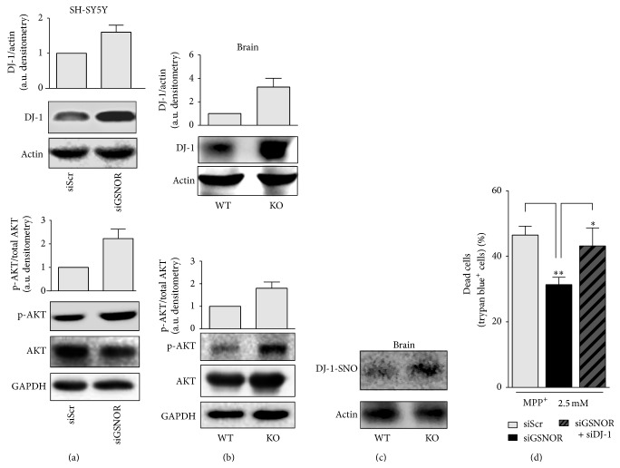
Figure 4.
DJ-1 and AKT in siGSNOR cells and GSNOR-KO brains. Western blot analyses of DJ-1, as well as basal and phosphoactive AKT in total extracts obtained from (a) siScr and siGSNOR SH-SY5Y cells or (b) GSNOR-KO and WT brains. Densitometric analyses of DJ-1 and phospho-AKT are shown on the top of the corresponding Western blot normalized to actin and basal AKT, respectively. (c) Biotin switch assay followed by pull-down with streptavidin and revealed with anti-DJ-1 antibody, in total extracts obtained from GSNOR-KO and WT brains. Western blot analysis indicates that DJ-1 was S-nitrosylated (present in the pull-down). Western blots shown are representative of at least n = 3 independent experiments that gave similar results. Actin or GAPDH were selected as loading controls. (d) Direct cell count upon Trypan blue staining of siScr and siGSNOR SH-SY5Y cells transfected or not with siRNA against DJ-1 (siDJ-1) and treated for 24 h with 2.5 mM MPP+. Graphs shown represent the mean of data ± SD of n = 3 independent experiments, ∗ P < 0.05 and ∗∗ P < 0.01.

