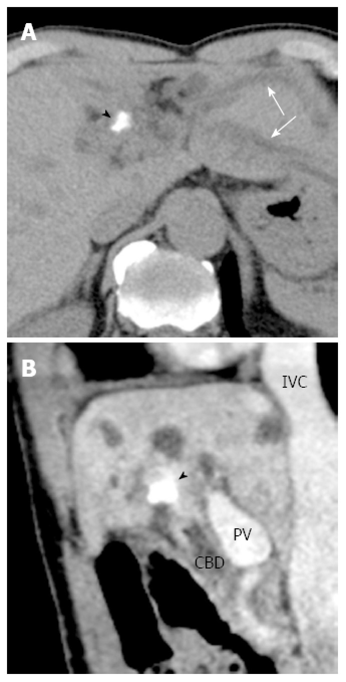
Figure 2.

Findings of computed tomography. A: Plain transverse computed tomography (CT) reveals a high-density area at the liver hilus (arrow head) with dilated left intrahepatic bile ducts (arrows); B: Sagittal plane of enhanced CT shows the perihilar high-density area (arrow head). PV: Portal vein; IVC: Inferior vena cava; CBD: Common bile duct.
