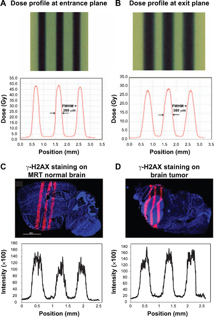
FIG. 2.
Microbeam profiles using Gafchromic films and γ-H2AX staining on irradiated brain. The beam width (FWHM) was 280 µm at the entrance plane on the top of the mouse brain (panel A) and 380 µm at the exit plane at the bottom of the head (panel B). Normal brain tissue section (panel C) and tumor-bearing mouse brain (panel D) were stained with anti-γ-H2AX antibody at 1 h after microbeam irradiation. Red fluorescence signal indicates the positive expression of γ-H2AX signal. Staining of γ-H2AX demonstrated the microbeam radiation track. The average FWHM of the γ-H2AX staining was 343 µm.

