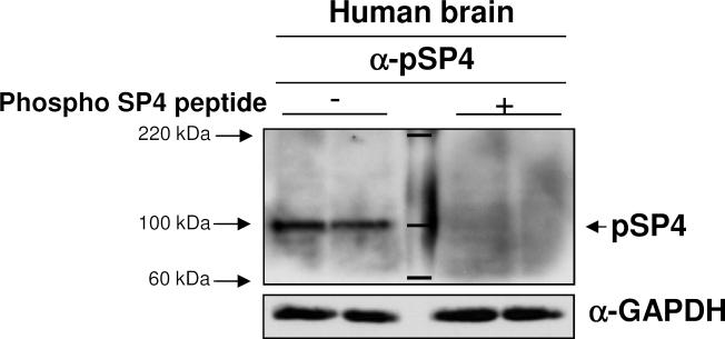
Figure 1. Expression of phosphorylated SP4 in human postmortem brain tissue.
Specificity of phospho-SP4 S770 antibody was determined by competition assay with peptide antigen in protein extracts from human postmortem cerebellum. Full images of immunoblots for phospho-SP4 S770 (left), phospho-SP4 S770 antibody co-incubated with peptide antigen (right) and GAPDH in postmortem cerebellum samples are shown from a representative bipolar disorder sample in duplicate. Total SP4 levels for this sample are shown in Figure 2A, since it corresponds to the fourth BD sample. Molecular marker weights are labeled and shown in kDa.

