
FIGURE 1.
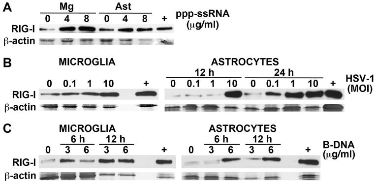
RIG-I protein expression in cultured primary murine astrocytes and microglia is upregulated following exposure to viral RNA and DNA motifs. Panel A: Cultured primary microglia (Mg) or astrocytes (Ast) were treated with transfection reagent alone (0) or transfected with uncapped 5′ppp-ssRNA (4 or 8 ug/ml). At 6 hrs following transfection RIG-I expression was assessed in whole cell protein isolates by immunoblot analysis. Panel B: Primary microglia and astrocytes were uninfected (0) or infected with HSV-1 (MOI of 0.1, 1, or 10). At 12 hrs following infection RIG-I expression was assessed in microglial whole cell protein isolates by immunoblot analysis and such expression in astrocytes was similarly assessed at 12 and 24 hrs. Panel C: Cells were treated with transfection reagent alone (0) or transfected with B-DNA (3 or 6 ug/ml). At 6 and 12 hrs following transfection RIG-I expression was assessed in whole cell protein isolates by immunoblot analysis. For comparison purposes, RIG-I protein expression in a similar number of resting HeLa cells is shown (+). Representative immunoblots are shown from one of three independent experiments.