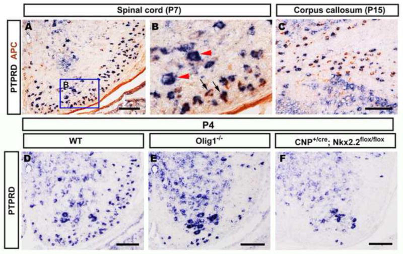
Figure 2.

Selective expression of PTPRD in differentiated oligodendrocyte lineage. A–C, P7 wild-type mouse spinal cord and corpus callosum sections were subjected to PTPRD ISH followed by anti-APC immunohistochemical staining. Double positive cells are represented by blue arrows; PTPRD positive only motor neurons are indicated by red arrowheads (B). D–F, P4 spinal cords from wild-type (D), Olig1−/− (E) and CNP+/cre;Nkx2.2fl/fl (F) were hybridized with PTPRD riboprobe. PTPRD expression in the white matter region was dramatically reduced in Olig1−/− and CNP+/cre;Nkx2.2fl/fl mutants. Scale bar: 100 μm.
