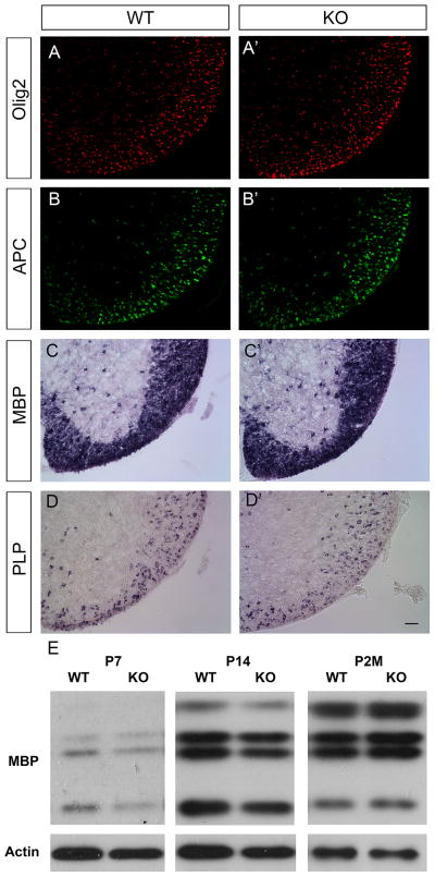
Figure 3.
Normal oligodendrocyte differentiation and myelin gene expression in PTPRD mutants. Traverse spinal cord sections from P7 wild-type (A, B, C, D) and PTPRD knock-out (A′, B′, C′, D′) littermates were subjected to immunostaining with anti-Olig2 (A, A′) or anti-APC (B, B′), and in situ hybridization with MBP (C, C′) or PLP (D, D′). Scale bar: 50 μm. E, Western immunoblotting of postnatal day 7, 14 and 2 months spinal cord extracts from wild-type and PTPRD mutants with anti-MBP and anti-β-actin antibodies. Comparable MBP expression was detected in PTPRD knock-out mice as compared to wild-type littermates.

