
Fig. 8. Effect of disulfide HMGB1 alone and /or in combination with prooxidants on TNF-α and IL-10 release in HEK-Blue mTLR4 cells.
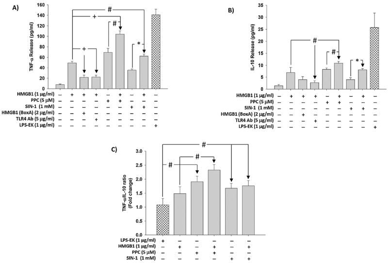
Cells were pre-incubated with HMGB1 (BoxA) or TLR4 pAb followed by stimulation overnight with HMGB1 in continued presence of the inhibitors. In the same set of experiments, cells were also incubated overnight with HMGB1, PPC or SIN-1 alone and/or in combination. Levels of TNF-α (Fig. 8A) and IL-10 (Fig. 8B) released into the culture medium were quantified using the ELISA kits of the relevant cytokine according to manufacturer’s instructions. The ratios of TNF-α to IL-10 (Fig. 8C) were calculated. The effect of LPS-EK on TNF-α and IL-10 release was used as positive control. The data represent 3 independent experiments. (#p ≤ 0.05, *p ≤ 0.01 and +p ≤ 0.001).