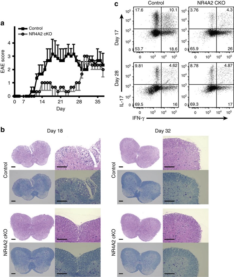
Figure 1. Mice lacking NR4A2 in CD4+ T cells are protected from early/acute EAE but develop late/chronic EAE signs.
(a) Clinical EAE score. Disease scoring for control NR4A2fl/fl (Control, black squares) and Cre-CD4/NR4A2fl/fl (NR4A2 cKO, grey circles). The mice with C57BL/6 background were immunized with MOG35–55 peptide emulsified in complete Freund’s adjuvant. Error bars represent s.e.m. (b) Histopathology of EAE. The spinal cords were fixed in formal-saline, paraffin embedded and sectioned before microphotography. Panels show adjacent sections from a representative mouse stained with haematoxylin and eosin (top row) or Luxol fast blue (bottom row) at × 4 (left) or × 10 (right) magnification; top panels are from a control mouse and bottom panels are from a NR4A2 cKO mouse; left panels are from day 18 post EAE induction, right panels are from day 32 post EAE induction from 1 of 2 independent experiments. Scale bars, 200 μm. (c) IL-17 and IFN-γ production by CNS CD4+ T cells as measured by intracellular cytokine staining following PMA/ionomycin stimulation in the presence of GolgiPlug for 5 h. The cells were isolated by enzymatic digestion of pooled CNS tissues during either early (Day 17) or late phase (Day 28) of EAE. Data are representative of at least three independent experiments, each using pools of three to five mice for each genotype and time point.

