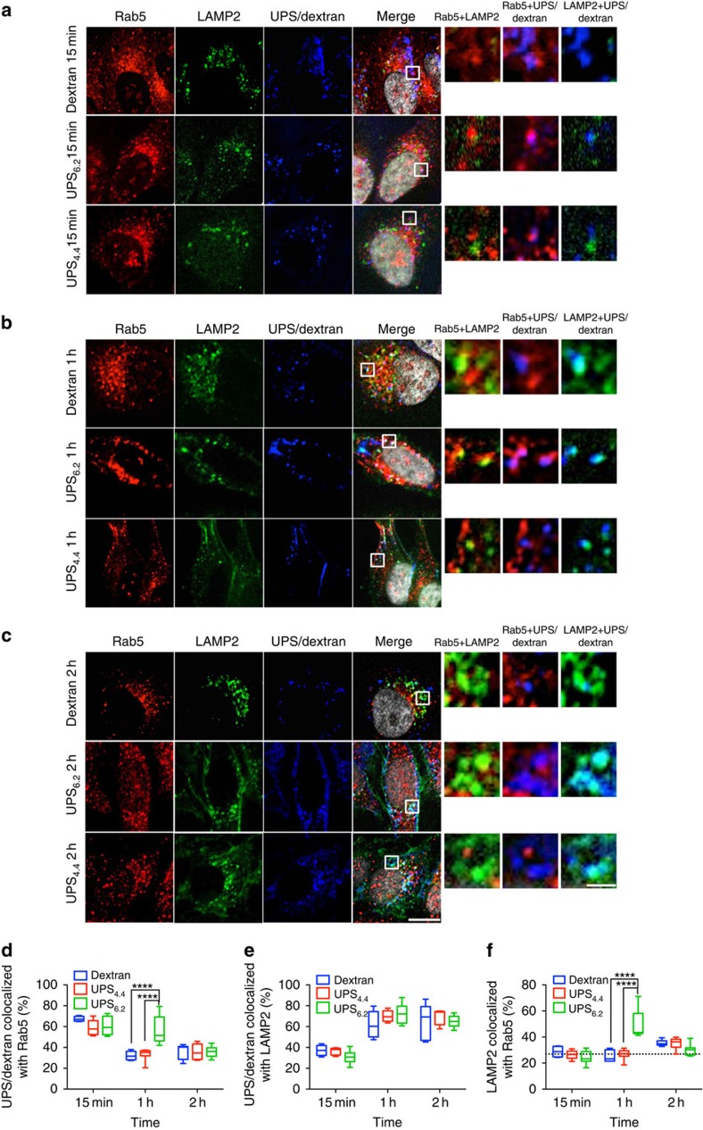
Figure 3. Buffering the pH of endocytic organelles affects their membrane protein dynamics.
HeLa cells were treated with 500 μg ml−1 dextran-TMR or 1,000 μg ml−1 UPS6.2-Cy5 or UPS4.4-Cy5 for 5 min for cell uptake. Then they were fixed after 15 min (a), 1 h (b) and 2 h (c). Immunofluorescence (IF) images show the localization of UPS nanoparticles in early endosomes (Rab5) or lysosomes (LAMP2). Scale bar, 10 and 5 μm (inset). Imaris software was used to analyse co-localization of z-stacked confocal images. The fraction of UPS/dextran co-localized with Rab5 (d) and LAMP2 (e) and the fraction of Rab5 co-localized with LAMP2 (f) were calculated from thresholded Mander's coefficient (see Supplementary Methods), n=10, α=0.05, ****P<0.0001. Two-way analysis of variance and Sidak's multiple comparison tests were performed to assess the statistical significance. The dashed line in (f) represents the basal level of Rab 5 and LAMP2 co-localization in HeLa cells without any treatment.

